Robotic SPECT enables intraoperative 3D tumor imaging
&width=710&height=710)
Molecular imaging
Tumor-targeting tracers that bind to prostate cancer cells are often administrated to patients to help identify disease. Depending on the clinical application, this tracer may be radioactive, fluorescent, or a combination of both.
After administration, tracers accumulate in prostate cancer lesions and/or in lymph nodes. Their distribution can subsequently be visualized using imaging modalities such as PET/CT and SPECT/CT. These large imaging gantries detect tracer-derived signals from outside the body and reconstruct them into three-dimensional images of tracer-avid tissues. The resulting images provide a roadmap for surgery by identifying the location of primary tumors and potential sites of metastatic disease, helping clinicians select suitable candidates for surgery and plan their excision of target tissues.
However, preoperative imaging does not always provide an accurate roadmap for identifying tumors during surgery. The acquired images represent a static snapshot obtained before surgery and may not fully reflect the intraoperative situation. During the procedure, tumor position can differ from that observed on the scan due to changes in patient positioning, respiratory motion, and displacement or manipulation of tissues and organs.
Surgical guidance
To localize tracers during surgery, surgeons rely on dedicated surgical guidance tools. One example is the drop-in gamma probe, previously developed at LUMC. When inserted into the patients abdominal cavity, this miniature detector detects radioactive signal emitted by tracer accumulated in the tumor or lymph nodes. When the probe approaches a suspicious lesion, the surgeon receives feedback in the form of acoustic and numeric signals. Another approach is the use of fluorescence, a concept in which specialized surgical cameras visualize fluorescent tissues, allowing the surgeon to identify tumors or sentinel nodes.
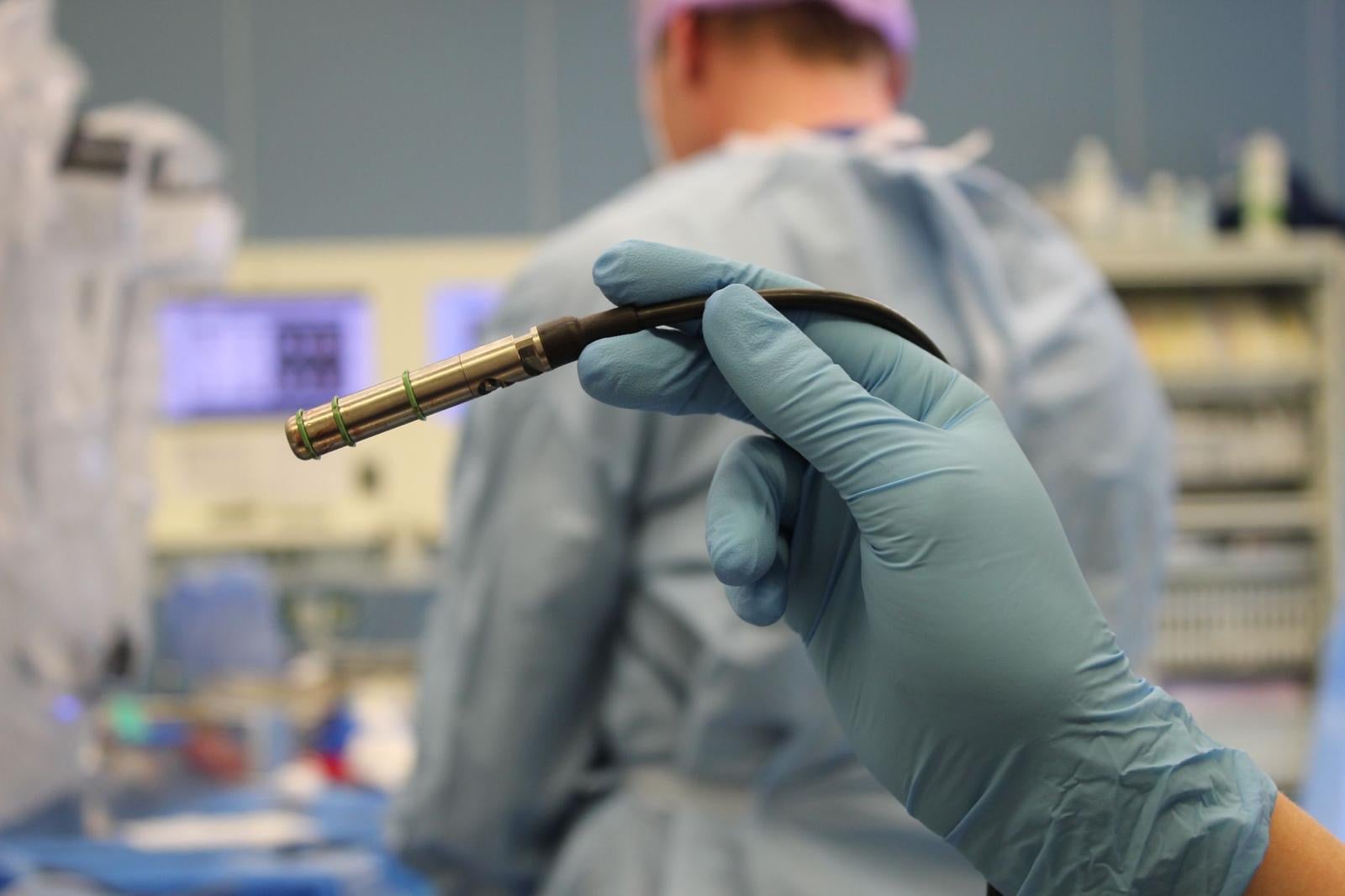
While these technologies provide valuable intraoperative guidance, both have limitations. A gamma drop-in probe does not provide a visual representation of the tumor itself. Also, the detected signal indicates that a tumor is present, but not how deep it is hidden within the tissue. Much like a metal detector looking for objects on the beach. Fluorescence on the other hand can only effectively identify a tumor when it lies near the surface. When there is overlying tissue the fluorescent signal becomes diffuse. Picture: drop-in gamma probe
An imaging system from within
To overcome the limitations of current technologies, researchers from the Interventional Molecular Imaging Laboratory at LUMC and the Department of Urology at the Netherlands Cancer Institute (NKI-AVL) developed Robotic SPECT. This novel technique converts the above-mentioned drop-in gamma probe detection into three-dimensional images of the tumor.
Physician-researcher Anne-Claire Berrens explains the underlying principle: “During surgery, the surgeon moves the drop-in gamma probe through the region where a tumor is expected. The probe continuously measures local radioactivity levels. At the same time, computer analysis of intraoperative video images determines the exact position and orientation of the probe within the abdominal cavity.”
By combining the probe’s position and orientation with the measured radioactive signal, the algorithm can calculate the location of the signal source within the body. “All measurements are then integrated into a three-dimensional image showing both the location and depth of a tumor or lymph node, effectively creating a robotic scan from within the body,” says Berrens.
Encouraging results
The researchers evaluated Robotic SPECT in 21 patients with prostate cancer, yielding encouraging initial results.
One group consisted of patients undergoing treatment for prostate cancer for the first time. In these patients, the aim was to identify sentinel nodes, the first lymph nodes to which cancer cells may metastasize. Robotic SPECT was compared with conventional SPECT/CT and fluorescence imaging. Robotic SPECT detected all affected sentinel nodes that had been identified on SPECT/CT, whereas fluorescence imaging missed 27% of these nodes. The advantage of Robotic SPECT was particularly evident for nodes located deeper within the tissue.
The second group consisted of patients with recurrent prostate cancer. In this setting, Robotic SPECT was compared with both PET/CT and SPECT/CT imaging. Robotic SPECT successfully identified all lesions that had previously been detected on PET/CT. Notably, conventional SPECT/CT, which is routinely performed in these patients, missed 71% of these lesions.
According to postdoctoral researcher Matthias van Oosterom, these findings can be explained by the nature of the new measurements. “With Robotic SPECT, measurements are acquired directly while placing the (drop-in) detector much closer to the source of radioactivity than a detector in a SPECT/CT scanner can be placed while located outside the body. This proximity improves signal detection and enables visualization of tumors missed on SPECT/CT.”
Anne-Claire Berrens highlights an additional advantage: “The Robotic SPECT can be adapted in real-time to the surgical situation. For example, measurements can be prolonged in an area of uncertainty, or the scan can be repeated if the surgeon requires additional information.”
Small device, broad potential
For now, the researchers are focusing on further development of the technology in this disease setting, but its potential applications are likely to extend far beyond prostate cancer. Robotic SPECT relies on radioactive signals to visualize tumors, a field that is evolving rapidly. “An increasing number of tumor-specific radiotracers are becoming available,” says Matthias van Oosterom. “Because Robotic SPECT measures and visualizes these signals during surgery independent of the compound they are connected to, the technique can thus be easily extended to other robot-assisted procedures.”
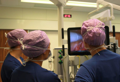 Another advantage is the compact dimensions of the system. Compared with conventional SPECT/CT gantries, the equipment is small, relatively affordable, and intuitive to use, which is likely to facilitate broader adoption across hospitals.
Anne-Claire Berrens emphasizes the clinical significance of the approach: “What was previously only a radioactive signal can now be translated into a 3D image of the tumor. This provides surgeons with improved environmental perception during the procedure, helping them decide where and how much tissue should be removed, with the aim of maximizing tumor resection while preserving healthy tissue wherever possible.”
Research in image-guided surgery
The results of this study were recently published in the European Journal of Nuclear Medicine and Molecular Imaging.
Over the past two decades, LUMC has played a leading role in the development of image-guided surgery using radioactive and fluorescent imaging technologies. These innovative approaches enable surgeons to visualize structures that are otherwise difficult to identify during surgery, including tumor tissue, nerves, sites of infection, and blood vessels. LUMC’s international standing in this field was recently highlighted in an independent bibliometric analysis of the scientific literature on fluorescence-guided surgery. The analysis ranked several LUMC researchers, including Alexander Vahrmeijer, Fijs van Leeuwen, Tessa Buckle, Nynke van den Berg, Cornelis van de Velde, and Matthias van Oosterom, among the world’s 20 most productive and influential authors in this research area, based on publication output and scientific impact.
More information about image-guided surgery research at LUMC can be found through:
- Green Light Leiden
- The Interventional Molecular Imaging Laboratory LinkedIn page
- The inaugural lectures of Professor Fijs van Leeuwen and Professor Alexander Vahrmeijer (both in Dutch)
Image-guided surgery research at LUMC can also be supported through a donation. More information is available via the LUMC Foundation.

&width=710&height=710)