Services
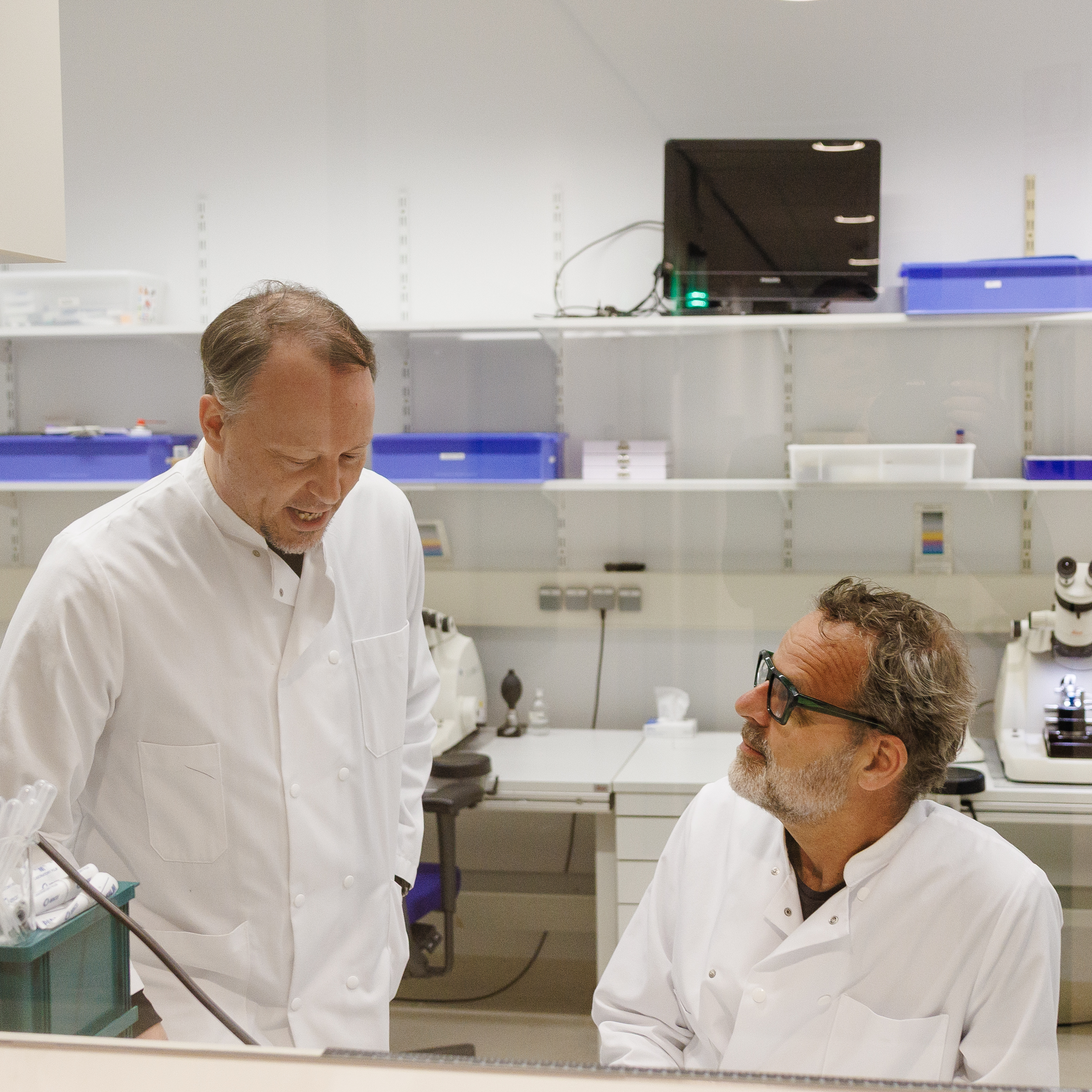
Consultancy
 Are you not sure what electron microscopy can do for you or what techniques to use?
We provide expert advice on the available electron microscopy techniques and will translate your question into a suitable and practical approach.
Training and Assistance
Training to use our specimen preparation instruments and electron microscopes is only available for experienced microscopists. After an introduction, assistance is available for technical issues. Beginner introductions or courses are not available.
Particle Analysis
Particle analysis involves the imaging of purified particles. Thes particles can be anything ranging from single proteins, protein complexes, protein aggregates (e.g. amyloid fibrils, microtubules), phages, viruses, virus like particles (VLPs) lipid-based nanoparticles (LNPs), extracellular vesicles (EVs), lipoproteins, organelles (e.g. lysosomes, mitochondria), nanoplastics, polymer nanoparticles.
These particles can be imaged in 2D or in 3D using the most appropriate technique, including negative stain EM, cryo-EM and scanning EM. (Note that single particle analysis cryo-EM is not part of our services.)
&width=400)
Cryo-EM image of an Extracellular Vesicle
&width=400)
Cryo-EM image of a liposome
&width=400)
Cryo-EM image of a Lipid Nano Particle
Cellular imaging
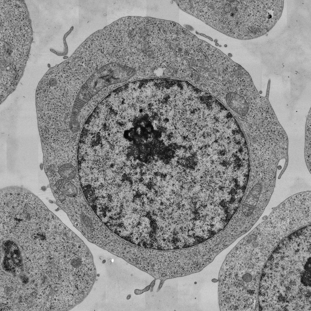 Cellular imaging typically comprises visualization of cellular morphology and the structural changes of cellular organelles, such as mitochondria, endoplasmic reticulum, lysosomes, endosomes, nuclei and also viral infections.
Imaging is typically performed on thin (100 nm) sections at 2 nm/pixel using automated large-scale 2D transmission electron microscopy, though 3D imaging using electron tomography, serial sectioning and 3D scanning electron microscopy are also available.
Organoid and Tissue Imaging
Imaging of organoids and tissues typically target the development and organization of different cell types, besides the visualization of subcellular structures.
Next to large scale 2D imaging, the method of choice is typically slice-and-view volume imaging through a block of plastic embedded material, which generates valuable information on organoid development and tissue health.
&width=710&height=710)
&width=710&height=710)
Protein Localization and Labeling
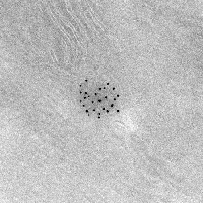 Localization or identification of certain proteins inside cells and tissues can be performed using antibody-gold labeling or correlative light and electron microscopy (CLEM) using antibody-bound fluorophores.
When protein labeling and fluorescent light microcopy does not provide enough resolution or clarity on the nature of underlying structures protein labeling can be combined with electron microscopy methods. Prior light microscopy imaging and good antibodies are desirable before starting EM labeling experiments.
Image Analysis and Visualization
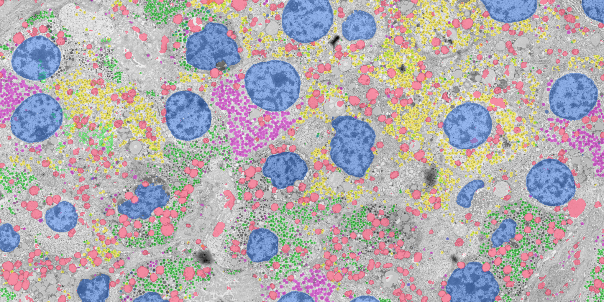 The analysis of images is always done together with the experts of our facility. The combination of biological and electron microscopy experts is imperative to draw scientific conclusions.
Image analysis also encompasses quantitative analysis of data. To perform objective analysis on large dataset we develop and use machine learning based segmentation of electron microscopy images, followed by quantification.
…The analysis of images is always done together with the experts of our facility. The combination of biological and electron microscopy experts is imperative to draw scientific conclusions.
Image analysis also encompasses quantitative analysis of data. To perform objective analysis on large dataset we develop and use machine learning based segmentation of electron microscopy images, followed by quantification.
At the electron microscopy department our primary output is the generation of images. We are experienced with the generation of images for publications, and also with the segmentation and visualization of three dimensional structures generated by tomography or 3D scanning electron microscopy.