Lab‑grown ‘mini‑inner ears’ shed new light on hearing loss
Peter Paul van Benthem, Wouter van der Valk and Heiko Locher
&width=710&height=710)
The findings were published in the scientific journal Nature Neuroscience.
The inner ear is invisible from the outside. Its location deep within the skull makes it hard to access. Many different conditions can cause hearing loss or balance problems, some of which arise very early in pregnancy. Until recently, these causes could barely be studied in humans. As a result, researchers do not know exactly know what causes these problems in humans. “For a long time, it was essentially a black box,” says researcher Wouter van der Valk. “We knew a lot was happening, but we couldn’t observe it properly.”
Grown from human stem cells
Van der Valk, group leader Heiko Locher, and department head Peter Paul van Benthem from the Department of Ear, Nose and Throat (ENT) at the LUMC have been cultivating mini‑inner ears in the lab for several years. These small clusters of cells, known as organoids, are made from human stem cells derived from skin or blood. By adding specific molecules step by step, scientists mimic the development of the inner ear as it occurs in an embryo.
The mini‑inner ears are only a few millimeters in size and far simpler than a real inner ear. Even so, they contain the key cell types found in the actual organ, including hair cells that detect sound and movement, and nerve cells that relay these signals to the brain.
One group of cells, however, had always been missing: the secretory cells known as dark cells in the vestibular organ and the stria vascularis in the cochlea. “These cells regulate the composition of the fluid in the inner ear”, Van der Valk explains. “Without them, hearing simply doesn’t work.”
New insights open the black box
For years, it was unclear how these secretory cells develop. The LUMC team therefore collected inner‑ear tissue from early, terminated pregnancies (first and second trimester), with donor consent and under strict ethical guidelines.
The researchers mapped the development of secretory cells in detail. “For the first time, we now have data from the human inner ear during early pregnancy”, says Van der Valk. “This gives us insights we never had before.”
They discovered that a signalling pathway called Hedgehog plays a crucial role. It acts as an on/off switch that determines how a cell develops. Their analysis showed that this signal must be switched off for the missing cells to appear. They tested this in the mini‑inner ears. “Once we turned off the Hedgehog signal, the previously missing cells appeared. Our inner ear model is now much more complete”, Van der Valk says.
This allows researchers to study hereditary hearing disorders far more precisely, as well as damage caused by viruses that attack the ear. It also enables more targeted testing of new treatments while significantly reducing the need for animal experiments.
What this means for patients
This research helps scientists and clinicians understand how the inner ear develops and what can go wrong in hearing loss or balance disorders. By creating a detailed developmental map of the human inner ear, researchers can better see how genetic abnormalities lead to problems with hearing or balance.
The mini‑inner ears also open new avenues for studying disease. They allow researchers to investigate not only hereditary hearing loss, but also damage caused by infections or harmful substances, such as certain medications.
The next step is testing new therapies in these models. “Think of gene therapy or RNA therapy, which may repair or bypass errors in genetic material. These models allow us to pinpoint which therapies are truly promising long before they reach patients”, Van der Valk says.
Tip of the iceberg
The LUMC is not the only centre developing inner‑ear organoids. The technique is also used in Boston, where the ENT researchers learned it and continue to collaborate closely. “We deliberately share our protocols with scientists worldwide so they can work with these models too”, Van der Valk says. The inner‑ear field is rapidly evolving, and the organ is only beginning to reveal its secrets. “We’re still at the very beginning,” he adds. “But we can already observe things that were previously impossible.”
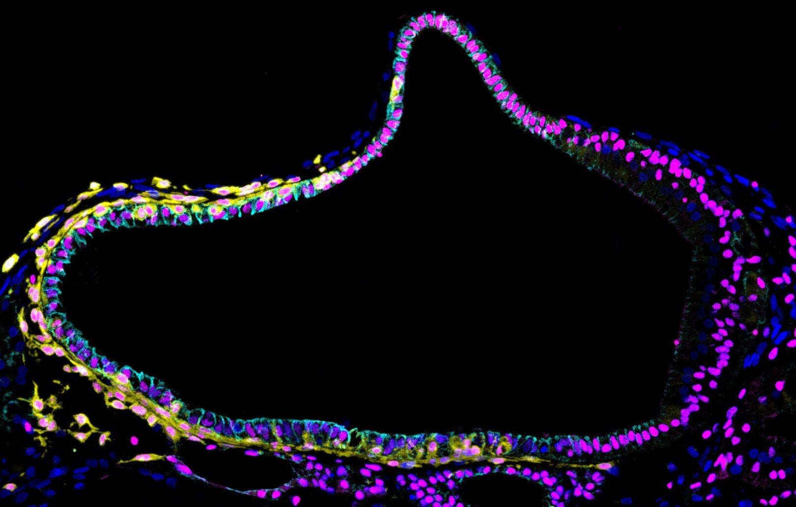
This is what a lab-grown mini inner ear looks like under a microscope.
- Shown in yellow: melanocytes (pigment cells)
- Shown in blue: epithelial cells, which line the inner ear
- And shown in pink: developing ear cells, among others.
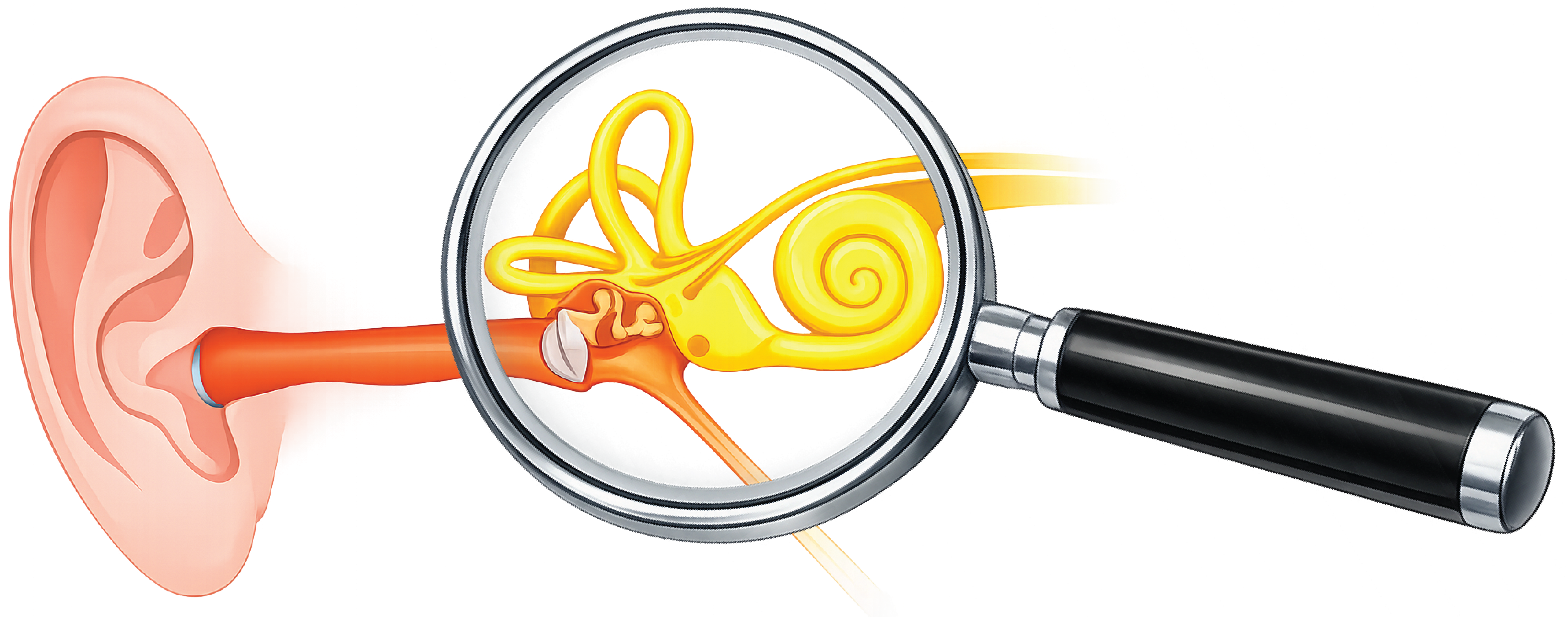
In the middle ear, the eardrum and the ossicles convert sound into fluid vibrations. In the inner ear, these vibrations are then converted into electrical signals that travel along the auditory nerve to the brain, where the sound is interpreted. The inner ear consists of the cochlea, the vestibular organ and the auditory nerve. It is invisible from the outside and difficult to access.
On 4 March, Wouter van der Valk obtained his PhD cum laude for his research on human inner‑ear development and inner‑ear organoids. His dissertation can be found here.

&width=710&height=710)
&width=710&height=710)