A heart that can defibrillate itself: LUMC and TU Delft work towards a shock-free future
&width=710&height=710)
Many heart rhythm disorders can be treated with medication or ablation. But these treatments do not work equally well for all patients. One example is atrial fibrillation, the most common heart rhythm disorder in the Netherlands, affecting around 300,000 people.
For some of these patients, an electric shock is sometimes the only way to restore a normal heart rhythm. This procedure is called electrical cardioversion. "The heart receives a strong electric shock that resets its rhythm. But because people remain conscious during an episode of an abnormal rhythm, a shock would be very painful and distressing", says Daniël Pijnappels, LUMC Professor of Cellular Electrophysiology. "That is why they have to go to hospital each time to undergo cardioversion under general anaesthesia."
People with atrial fibrillation therefore cannot simply be treated with an ICD, a device that delivers an electric shock to stop dangerous heart rhythms while a person is awake.
Letting the heart defibrillate itself
Our heart continuously produces electrical signals. Doctors currently use these signals mainly to diagnose heart problems, for example with an ECG. "We want to use the electrical signals that the heart naturally produces to stop the abnormal rhythm as well", says Pijnappels.
The solution the team is developing for this and other groups of patients is technically complex, but, according to Pijnappels, also "surprisingly logical". "The heart functions through electrical signals. By controlling these signals, you can influence the heart rhythm and make the heart defibrillate itself", he says.
The team has developed this idea into a biologically integrated cardiac defibrillator: the BioICD.
New biology: making the heart sensitive to light
The BioICD is equipped with LEDs that use flashes of light to generate the electrical signals needed to influence and restore the heart rhythm. First, however, the heart needs to acquire an ability it does not naturally have: responding electrically to light.
Using gene therapy, the researchers introduce special proteins into heart cells that enable them to do this. When light hits the cells, they produce a small electrical signal. "This allows us to control very precisely where, when and for how long a heart cell produces an electrical signal, and how strong that signal is", Pijnappels explains. "This helps us determine how much electrical stimulation is needed to stop an abnormal rhythm."
New technology: smart LED systems
A heart that responds to light also needs a light source that can be activated at exactly the right time and in exactly the right place. This is where the collaboration between LUMC and TU Delft comes in. Together, the researchers have developed small, smart LED systems that can be implanted deep inside the body and deliver precisely the right amount of light at the right moment.
"We can implant these LED systems in animals", says Pijnappels. "We have now demonstrated the principle in rats. The next step is research in pigs." The technology is not yet ready for use in humans, but he says the prospects are promising.
Human heart tissue grown to the size of a real heart
To understand how a flash of light can stop an abnormal heart rhythm, the researchers first need to understand exactly how heart cells respond. They cannot simply test this in humans. Instead, they use a safe intermediate step: a kind of artificial heart made from real human tissue grown in the laboratory.
The researchers take human heart cells and grow them into a large layer of tissue in the laboratory. This layer is approximately the size of the atrial tissue in a human heart. "Size matters when studying heart rhythm disorders", says Pijnappels. "Heart cells behave differently in a small piece of tissue than they do in a model that is closer to the size of the real heart."
In this model, the researchers can induce abnormal rhythms, test different light patterns and observe how the rhythm changes when the LEDs are activated.
Donated heart tissue
The heart cells used by LUMC come from donated human heart tissue. Within the laboratory, assistant professor Twan de Vries works on developing and growing these cells. This allows the cells to be used repeatedly for research. In science, such a continuously growing population of cells is known as a cell line. LUMC has unique human cell lines that may be of interest for both academic and commercial research.
Stopping an abnormal rhythm with a smartwatch
The researchers have also been looking for ways to activate the LED system when an abnormal heart rhythm occurs. This led to the idea of a smartwatch that detects an abnormal rhythm and sends a signal to a small receiver under the skin. The receiver could then activate the small, smart LED system attached to the heart.
"You put your smartwatch on your chest and your heart restores its own rhythm. Without you feeling any pain", says Pijnappels, placing his wrist against his chest. It sounds futuristic, and such a smartwatch does not yet exist. Before this technology could ever be used in humans, extensive testing would be needed.
Fully biological defibrillation
The BioICD itself also needs to be tested, refined and validated further. The technology still has a long way to go. But if everything works as hoped, this approach could fundamentally change the treatment of heart rhythm disorders.
"The ultimate goal is to make the heart detect and stop an abnormal rhythm itself. Without electronics or light, but fully biologically and without a shock. The heart as its own defibrillator", says Pijnappels.
A laboratory where disciplines come together
The heart is a complex organ where biology, physics, chemistry, engineering and medicine come together. That is why doctors, biologists, engineers, physicists and research analysts work side by side in the Laboratory of Experimental Cardiology, led by Pijnappels.
The laboratory is designed so that different types of research can literally take place side by side: from pipetting to computer simulations and soldering. This allows researchers and students from different disciplines to work together and learn from one another. "Our goal is to bring biology and technology together so that we can better understand and treat heart disease", says Pijnappels.
Among other things, the team wants to understand what goes wrong when heart rhythm disorders occur and develop innovative solutions to treat them.
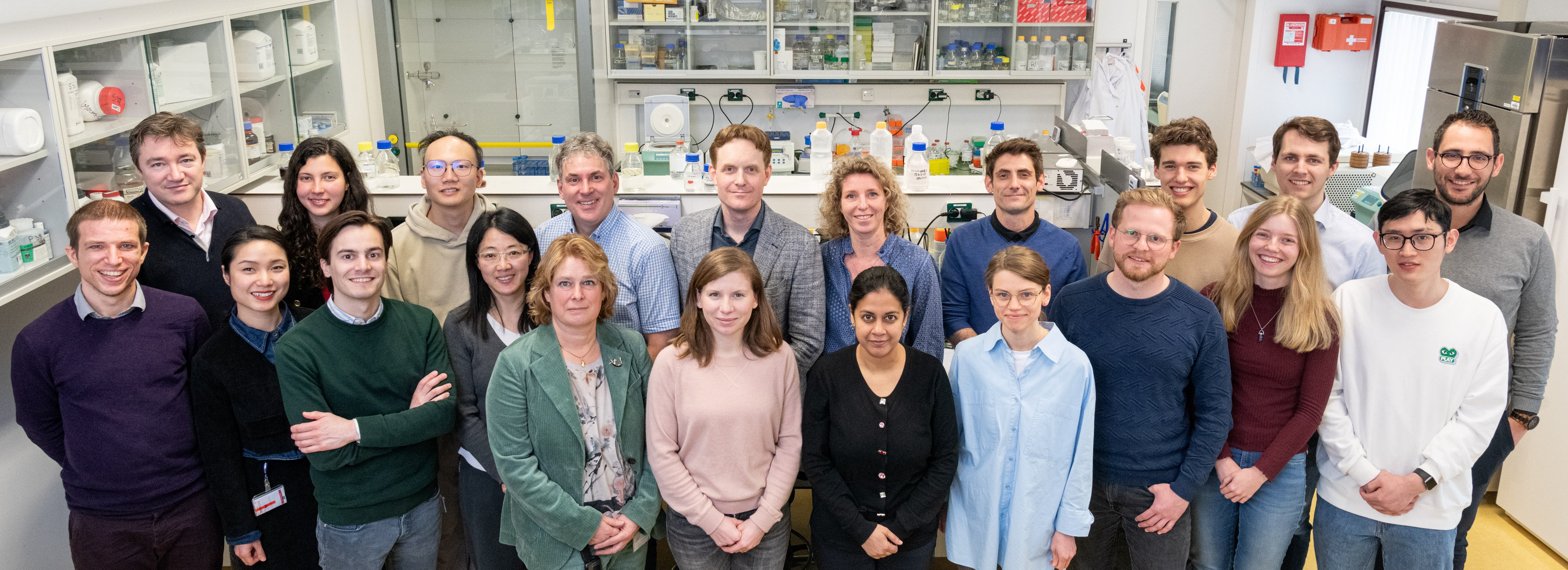
Researchers at the Laboratory of Experimental Cardiology.
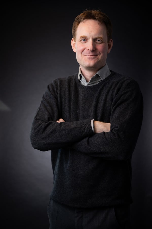
Daniël Pijnappels is the head of the Laboratory of Experimental Cardiology and a professor of cellular electrophysiology at LUMC. He is also a Medical Delta professor at Delft University of Technology (TU Delft), in the Department of Microelectronic Engineering within the Faculty of Electrical Engineering, Mathematics, and Computer Science (EWI). In addition to being a researcher, he also considers himself an inventor.
&width=710&height=710)
&width=710&height=710)
&width=710&height=710)