Image guided surgery
&width=826&height=464)
Benefit for patients
By improving individual aspects of surgery, the LUMC aims to increase the balance between surgical outcome and surgical side effects. By improving intraoperative tumour detection and by providing a better delineation of the disease, the number of radical resections can be increased. Image guided surgery techniques can also make the surgical procedures safer by minimizing damage to healthy tissue. Patient benefits related to technologies that help minimise the surgically induced side-effects may be found in factors such as: preservation of function, reduced surgical morbidity, and more rapid reintegration into society.
…Benefit for patients
By improving individual aspects of surgery, the LUMC aims to increase the balance between surgical outcome and surgical side effects. By improving intraoperative tumour detection and by providing a better delineation of the disease, the number of radical resections can be increased. Image guided surgery techniques can also make the surgical procedures safer by minimizing damage to healthy tissue. Patient benefits related to technologies that help minimise the surgically induced side-effects may be found in factors such as: preservation of function, reduced surgical morbidity, and more rapid reintegration into society.
Benefit for imaging-agent, soft- and hard- ware developers
In addition to driving independent academic research projects, the LUMC offers a unique combination of extensive expertise and research infrastructure to external private and public partners who are pursuing the development of imaging technologies for image guided surgery applications. At any point in the development trajectory we can offer our expertise for a fast, high quality translation. This support spans from technology/intellectual property development (e.g. chemistry, modalities, augmented- virtual-reality concepts) to preclinical and clinical technology evaluation.
Benefit for surgeons
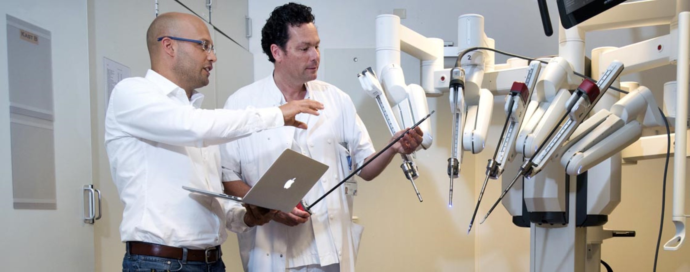
The value of image guidance technologies is increasing with the continuous move towards minimally invasive surgical procedures, in which the surgeon operates through small incisions using a laparoscope and specialised instruments. Here the surgeon cannot rely on touch to distinguish between the tumour and healthy tissue. Using a tissue specific imaging agent, possibly coupled with a tailored navigation approach, can help the surgeon to operate with more confidence around delicate structures.
Two internationally acknowledged LUMC research groups have as main focus image guided surgery
The Interventional Molecular Imaging laboratory (Radiology) is focusing (together with its industrial and academic partners) on the development, translation and implementation of molecular imaging technologies that support image guided interventions. These efforts are coupled to educational and training activities.
The group of Dr. Alex Vahrmeijer (Surgery) is focussing on precision surgery to improve the treatment of cancer patients. In a unique collaboration involving LUMC, CHDR and national and international partners current research focuses on the introduction of real-time tumour detection and normal structure delineation using both clinically available probes and novel tumour specific probes.
The impact of the work performed these groups is not only reflected by a high number of scientific publications, and key positions in the organization structure of relevant academic societies and the European Association of Nuclear Medicine and Molecular Imaging, but also in the abundance in competitive personal scholarships obtained on the topic of image guided surgery e.g. VENI-, VIDI-, and VICI- grants awarded by the Netherlands Organisation for Scientific Research, multiple ERC-grants and multiple personal KWF scholarships, regular KWF Grants and NIH funding.
Dr. Fijs van Leeuwen
Radiology
&width=826&height=464)
Dr. Alex Vahrmeijer
Surgery
&width=826&height=464)
Image guided surgery can be applied to several different scientific and clinical fields.
Targeted tissue removal
In a unique collaboration involving the LUMC and national and international partners, the image guided surgery research efforts at the departments of Surgery and Radiology focus on the development and clinical introduction of new technologies that can advance patient care.
…Image guided surgery can be applied to several different scientific and clinical fields.
Targeted tissue removal
In a unique collaboration involving the LUMC and national and international partners, the image guided surgery research efforts at the departments of Surgery and Radiology focus on the development and clinical introduction of new technologies that can advance patient care.
By their own merit or together with their partners, the LUMC-based research groups deliver high quality solutions that help to visualize surgically relevant lesions. Combining our longstanding expertise in chemistry, engineering, (pre-) clinical validation, and an efficient infrastructure, we offer a one-stop-shop approach for fast clinical translation of new technologies . In this process we support knowledge transfer, education and training.
Most of the efforts towards the targeted removal of diseased tissues focus on oncological applications in oncological surgery (e.g. pancreatic, colorectal, brain, lung, breast, head-and-neck cancer, gynaecology, urology, dermatology and orthopaedic indications). Besides this alternative image guidance applications are being pursued in for instance interventional radiology and infectious diseases.
Five key examples of technologies that have been developed in-house and have found their way to the clinic are:
- The hybrid tracer Indocyanine green (ICG) ICG-99mTc-nanocolloid for sentinel node identification; Commercially available and currently used in routine surgical care.
- The DROP-IN gamma probe for robot-assisted radioguided surgery; Currently evaluated during sentinel node procedures and PSMA-targeted salvage surgery
- Multispectral fluorescence imaging of multiple fluorescent contract agents; Anticipating the future availability of imaging tracers for different targets the concept of multispectral imaging has been evaluated in different settings.
- Hybrid surgical guidance modalities that detect both fluorescent and radioactive signatures; A variety of hybrid surgical guidance technologies have been studied in clinical trials, one of which is currently commercially availableReal time detection of colorectal liver metastases with ICG as standard of care
- Real time detection of ureters during surgeries in the lower pelvis and during kidney transplant procedures with newly designed agents…
Four key examples of technologies developed by external partners that have undergone clinical evaluation are:
- First-in-human evaluation of EM-137 (at the time GE-137); This c-Met targeted tracer has been evaluated for its ability to target gastrointestinal cancers and head and neck cancer
- Evaluation of tissue perfusion using hyperspectral imaging
- First-in-human evaluation of OTL-38 for the detection of ovarian cancer.
- Clinical implementation of augmented and virtual reality navigation concepts during soft-tissue surgery in head and neck cancers and urological applications.
Sparing of critical anatomical structures
In some cases the side effects as result of damage being surgically inflicted to delicate vital structures can be quite severe, meaning that the patients could be free of the disease, but would still persist to have serious health complaints. Especially, the elderly may benefit from less invasive procedures as they are prone to experiencing the side effects of for instance pelvic surgery e.g. leading to pelvic organ dysfunction such as urine and faecal incontinence and sexual dysfunction. We reason that implementation of precision surgery applications will could help increase the Quality of Life of patients significantly.
Imaging technologies that enable surgeons to reduce intraoperative injury, or just help preserve specific structures, may be seen as a true “game changer”. Here different routes can be pursued: 1) preoperative identification of e.g. nerves so that the chance of surgical side effects can be estimated and 2) the development of tracers specific for delicate anatomical structures. We explore both routes.
Next to minimisation of damage to the lymphatic anatomy, and ureter, nerve sparing surgery is one of the key focus areas. Peripheral nerve injury presents a notable (surgical) complication and can be very debilitating for patients; 5% of patients that undergo surgery have post-surgical morbidity that can be related to intraoperatively induced peripheral nerve injury.
Collaborations
The two research groups work closely together with other departments within the LUMC. Especially our GMP facility is important in probe development and clinical validation.
Also the groups have long-term collaborative research alliances with leading institutes and consortia around the world. In addition to classic academic research institutions intensive collaborations exist with non- academic representatives, such as commercial enterprises, for example in the field of probe and camera development.