Groundbreaking technique reveals brain's smallest blood vessels for the first time
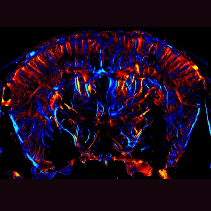
Ultrasound image of a mouse brain. The colors show how fast small particles move through blood vessels in the brain tissue: blue means upward, red means downward.
&width=710&height=710)
With current medical imaging techniques like MRI, CT scans, or standard ultrasound, only the larger blood vessels are visible. Yet, 80% of our circulatory system consists of capillaries: extremely small vessels with a diameter as tiny as 5 micrometers (0.005 millimeters), which, until now, remained invisible. "There was no device capable of visualizing the smallest blood vessels deep within organs. The best technology available could only zoom in up to 0.1 millimeters", says lead researcher Franck Lebrin.
Now, the researchers have developed a device that allows high-resolution imaging of the brain's tiniest capillaries, not just their structure, but also their function: how blood flows, how the vessels respond, and whether abnormalities are present. "This is a real revolution", says Lebrin. "For the first time, we can observe what's happening in the brain's smallest blood vessels. This opens the door to earlier diagnosis, better treatments, and perhaps even prevention of vascular diseases."
The findings have been published in the journal Nature Biomedical Engineering.
Transporting oxygen
Blood vessels carry blood throughout the body: delivering oxygen-rich blood from the heart to organs and tissues and returning oxygen-poor blood to the heart and lungs. Capillaries play a crucial role by supplying oxygen and nutrients directly to cells, enabling them to function properly and stay healthy.
Sometimes, however, capillaries malfunction. As a result, certain brain areas may not receive enough oxygen, harmful substances can enter, and chronic inflammation may develop. This can lead to irreversible brain damage that causes dementia. Unfortunately, symptoms often only appear once the damage has progressed too far to be treated.
95% of brain vascular map now complete
 Yet these abnormalities often begin in the capillaries up to twenty years before symptoms arise. With this new device, researchers can now observe what goes wrong in the earliest stages, opening up new possibilities for earlier detection and testing of new medications.
The technique is based on a highly advanced form of ultrasound. According to Lebrin (photo right), the device is more detailed than ever. "Imagine the brain’s blood vessels as a road network: previously, we could only see the highways. Now we can also see secondary roads, streets, and country lanes. We've jumped from 20% to 95% visibility in one leap", says Lebrin, who believes that within a few years, it will be possible to visualize the remaining 5% of currently invisible capillaries as well.
Successfully tested
The system works like a type of helmet placed on the head, while a receiver scans the brain. No surgery is required. Scientists therefore describe the method as non-invasive. It was first tested in the lab on mice with hereditary hemorrhagic telangiectasia (HHT), a rare genetic disorder involving fragile and dilated capillaries prone to bleeding. The cause often lies with pericytes, cells that normally stabilize small blood vessels and regulate blood flow and the blood-brain barrier. When they malfunction, problems arise.
The researchers created detailed images showing exactly how vessels change in the disease, how blood flows, and how abnormal vascular connections form. Since the smallest blood vessels in mice and humans are the same size, Lebrin expects the results to translate well to humans.
Study with potential new drug
At the end of this year, the new imaging device will be used to study patients with RVCL-S, a rare hereditary small vessel disease, in collaboration with the LUMC Neurology Department. Simultaneously, Lebrin's research group will begin testing new self-developed drugs targeting pericytes. The device will allow them to directly measure the drug's effect on the brain’s vasculature. The participating patients have not yet developed symptoms but do carry the genetic mutation for RVCL-S or HHT. This allows intervention at a very early stage.
A technique with broad potential
The research starts with HHT, but according to Lebrin, the device's potential is much broader. The technique could also be applied to other organs such as the heart, kidneys, and beyond. LUMC is already using it in organ donation to assess whether a donor kidney is well-perfused. In the future, the technology could help detect other diseases associated with vascular abnormalities at an early stage.
From lab to clinical practice
This study* is a collaboration between LUMC and the Institute of Physics for Medicine in Paris (part of the French Inserm institute), which developed the clinical system. The partnership has led to the recent creation of an International JointLab and the founding of a spin-off company focused on pericyte-based treatments.
* The research project, named MICROVASC, is funded by the European Innovation Council (EIC) under the EU’s EIC Pathfinder Challenge.

&width=710&height=710)
&width=710&height=710)
&width=710&height=710)