Jongbloed Group - Morphology and Development of Congenital Heart Disease
The first pilar, Basic science. Aimed at mechanisms and sequelae of congenital/structural heart disease, in particular arrhythmias. Special emphasis is on cardiac autonomic innervation, as this drives the conduction system and its role in arrhythmogenesis is increasingly recognised. Arrhythmias related to pathological cardiac innervation patterns due to disturbed epicardial signalling are currently the groups’ main focus. The group uses in vivo and vivo animal models as well as human tissues.
…The first pilar, Basic science. Aimed at mechanisms and sequelae of congenital/structural heart disease, in particular arrhythmias. Special emphasis is on cardiac autonomic innervation, as this drives the conduction system and its role in arrhythmogenesis is increasingly recognised. Arrhythmias related to pathological cardiac innervation patterns due to disturbed epicardial signalling are currently the groups’ main focus. The group uses in vivo and vivo animal models as well as human tissues.
The second pilar, Clinical and anatomical science. aimed at phenotyping the morphological spectrum of congenital heart disease as related to disease outcome, such as arrhythmias. These studies take advantage of the unique Leiden Collection of Congenital Malformations (tpart of the Biobank Congenital Heart Disease) at the dept. of Anatomy & Embryology, as well as of data of clinical care paths (dept. of Cardiology, Heart Lung Center). A special project is the development of dedicated 3D printing models of congenital heart diseases.
Reseach focus
Congenital heart disease (CHD) is the most common birth defect, affecting approximately 1 in 1,000 live-born children. Over recent decades, therapeutic advances have dramatically improved survival rates, allowing increasing numbers of patients to reach adulthood. However, despite improved early survival, late complications remain a significant concern. These include life-threatening arrhythmias that can lead to sudden cardiac death (SCD), particularly in high-risk subgroups such as patients with right ventricular dysfunction and those with Fontan circulation.
…Congenital heart disease (CHD) is the most common birth defect, affecting approximately 1 in 1,000 live-born children. Over recent decades, therapeutic advances have dramatically improved survival rates, allowing increasing numbers of patients to reach adulthood. However, despite improved early survival, late complications remain a significant concern. These include life-threatening arrhythmias that can lead to sudden cardiac death (SCD), particularly in high-risk subgroups such as patients with right ventricular dysfunction and those with Fontan circulation.
Understanding cardiac morphology and phenotypic variations is essential for predicting and managing these complications. Moreover, identifying the molecular mechanisms underlying structural changes in cardiac tissue is crucial for potentially intervening in disease processes triggered by hemodynamic overload or ischemia.
Among acquired heart diseases, myocardial infarction (MI) remains a leading cause of death in developed countries. In MI survivors, sudden cardiac death has been linked to abnormal innervation patterns in the left ventricle, resulting in autonomic hyperinnervation. This creates electrophysiological instability that can trigger fatal arrhythmias. Accumulating evidence demonstrates a clear relationship between ventricular arrhythmias, sudden cardiac death, and sympathetic nervous system activity.
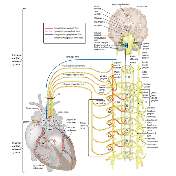&width=710 "Figure 1 - The cardiac autonomic nervous system.")
Figure 1 - The cardiac autonomic nervous system.
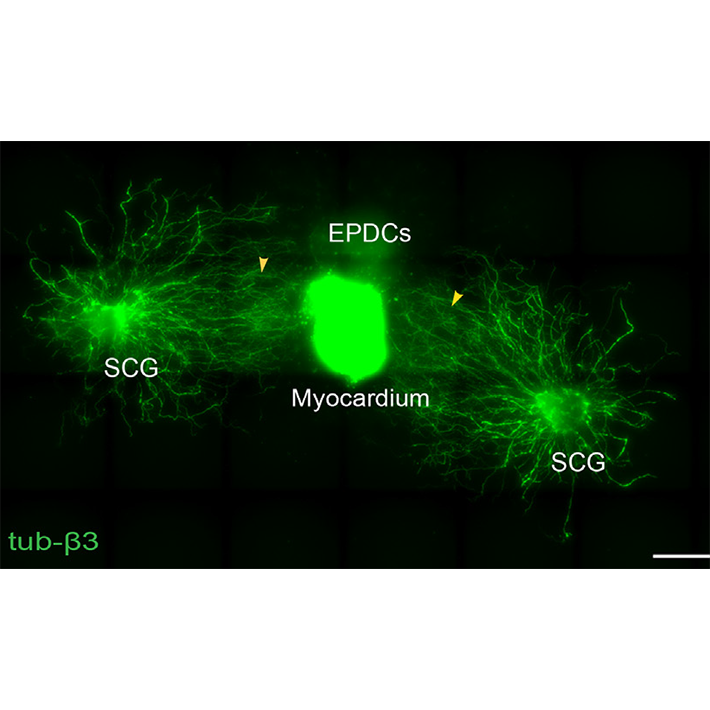&width=710 "Figure 2- Ganglion cultures")
Figure 2- Ganglion cultures
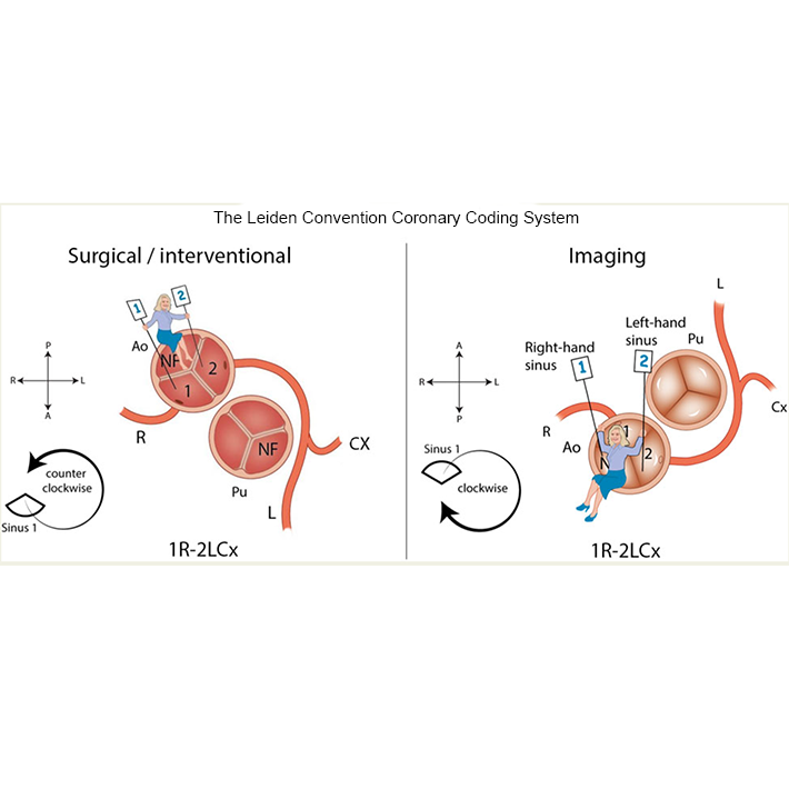&width=710 "Figure 3 - The Leiden Convention Coronary Coding system")
Figure 3 - The Leiden Convention Coronary Coding system
Clinical anatomy-3D printing of congenital malformations
A special project is the development of dedicated 3D printing models of congenital heart diseases for educational and procedural planning purposes, that are developed on the dept. of Anatomy & Embryology (Figure 4). The complex anatomy, treatments and risk of late complications may have an important impact on the life of congenital heart disease patients. Adequate knowledge transfer is very important for disease insight and understanding, not only for the patient, but also for his/her family. This project is aimed at improving information for patients with congenital heart defects and their family members through 3D printing. We have developed protocols for creation of detailed colour 3D prints of congenital heart defects, based on CT and MRI scan of patients. In the past years, several 3D printing models of congenital heart defects have been developed, that are used in the consultation room for patient education.
…A special project is the development of dedicated 3D printing models of congenital heart diseases for educational and procedural planning purposes, that are developed on the dept. of Anatomy & Embryology (Figure 4). The complex anatomy, treatments and risk of late complications may have an important impact on the life of congenital heart disease patients. Adequate knowledge transfer is very important for disease insight and understanding, not only for the patient, but also for his/her family. This project is aimed at improving information for patients with congenital heart defects and their family members through 3D printing. We have developed protocols for creation of detailed colour 3D prints of congenital heart defects, based on CT and MRI scan of patients. In the past years, several 3D printing models of congenital heart defects have been developed, that are used in the consultation room for patient education.
Several of these models can be found at: https://anatomytool.org/content/congenital-heart-diseases
In addition, personalised print are used for procedural planning, such the treatment of aortic coarctation by stenting (Figure 5).
The project is part of the activities of the Expertise Center for Cardiovascular Development and Disease (ECCARD).
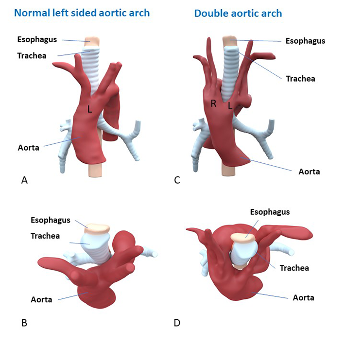&width=710 "Figure 4 - 3D printing of double aortic arch")
Figure 4 - 3D printing of double aortic arch
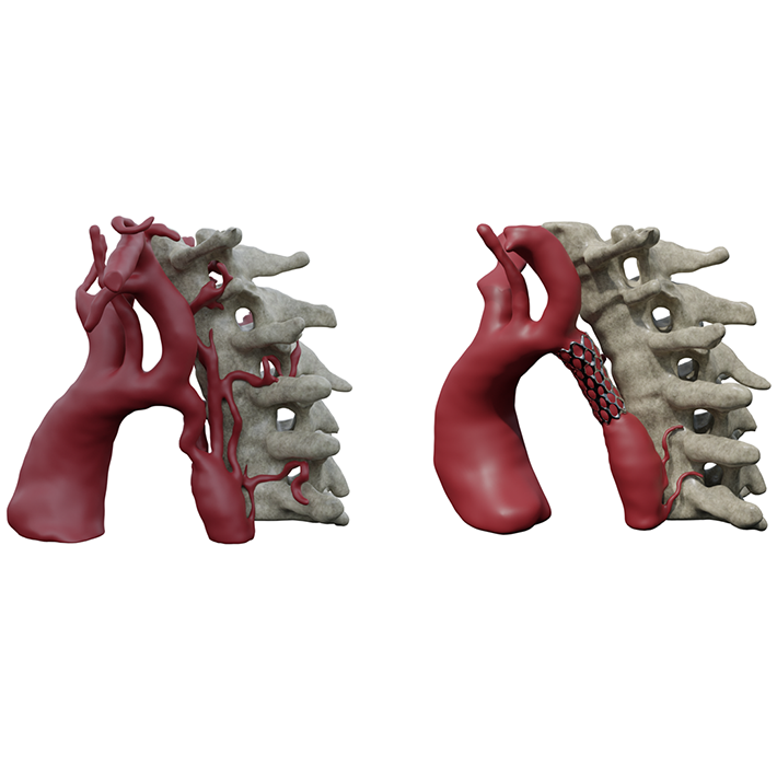&width=710 "Figure 5 - 3D Print coarcatio aortae. Left: pre-procedural, right: after percutaneous stenting of the coarctation.")
Figure 5 - 3D Print coarcatio aortae. Left: pre-procedural, right: after percutaneous stenting of the coarctation.
Themes for innovation and Societal Outreach
Key publications
Our team
- Prof. Dr. Monique Jongbloed – Cardiologist/ Full Professor, group leader
- Conny van Munsteren – research technician
- Michiel Blok – researcher
- Thomas Bos – MD PhD candidate
- Lieke van Roon – PhD candidate
- Liza Polyokova – PhD candidate
- Eline Mol – PhD candidate