De Ruiter Group - Clinical anatomy and cardiovascular development
To provide better mechanistic insights in the pathomorphology of patients our human studies are paralleled by studies in well-defined mouse models to unravel the role of extracardiac contributors (second heart field, neural crest and epicardium) in the differentiation and maturation of the aortic root. The analyses include a range of immunohistochemistry and molecular biological approaches (ao single cell RNA sequencing), in combination with (genetic cre-lox system and non-genetic fluorescent dye microinjections) cell lineage tracing techniques, genetic models for bicuspid aortic valves (Nos3 knock out mouse model) and transpostion of the great arteries (PDGFRalpha ko), laser dissection microscopy, in vitro flow experiments, electrophysiology, optical mapping and ultrasound evaluations of embryonic cardiac function.
With these techniques we explore the hypothesis that processes essential for normal embryonic cardiovascular patterning and differentiation are also involved in the maintenance and repair of the heart and vessel wall. Small deviations in the signaling processes can result into congenital cardiovascular malformations seen perinatally, but also into increased susceptibility of cardiovascular pathology seen in adults.
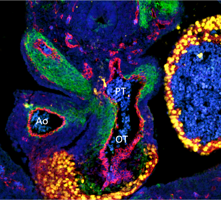
Figure 1 - Cell lineage in the outflow tract of the heart. The green cells are from the neural crest and contribute to the vascular wall of the Aorta (Ao) and the pulmonary trunk (PT). In red are the endothelial cells covering the inner side of the heart and bloodvessels. The red cells with yellow nuclei are the myocardial cells of the cardiac outflow tract (OT).
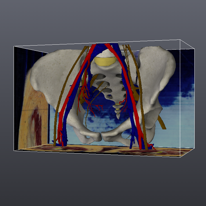
The Section of Clinical Anatomy has a consultant function in surgical research. We support, supervise and/or coordinate research in close collaboration with clinical departments on new relevant neuroanatomical, orthopedic, abdominal and pelvic surgical scientific questions and surgical procedures. A major part of these studies concerns the development of nerve preservation techniques to reduce postoperative morbidity after complex surgery. For our long-term objective to overcome surgical problems we are developing a unique 3D surgical planning system based on high resolution digitized histological sections of the human.
3D: OAH webviewer weblink: http://anatomy.tudelft.nl
Themes for innovation and Societal Outreach
Key publications
Our team
- Prof. Dr. Marco de Ruiter – full professor, group leader
- Dr. Margot Bartelings – assistant professor
- Bert Wisse – senior research technician
- Ruben Methorst – PhD candidate
- Tamara Borsboom – PhD candidate