LUMC and TU Delft help build the largest brain atlas ever created: first results published
Researchers from the Department of Radiology (from left to right): Baldur van Lew, Thomas Kroes, Jeroen Eggermont, Boudewijn Lelieveldt, Soumyadeep Basu, Julian Thijssen, Chang Li, and Alexander Vieth.
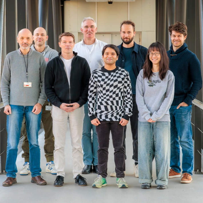
Researchers from Leiden University Medical Center (LUMC) and Delft University of Technology (TU Delft), through the Medical Delta collaboration, developed the software behind the atlas. The tool allows researchers to navigate through the atlas and map cells and their characteristics. “We turned an ocean of search possibilities into a bathtub,” says principal investigator and professor Biomedical Imaging Boudewijn Lelieveldt.
The first part of the atlas has now been published in the scientific journal Cell. In total, the project consists of 26 scientific articles. Dutch researchers made significant contributions to six of them.
The project is led by the Allen Institute in Seattle, but researchers from across the globe are involved. Their goal is to map all approximately 200 billion cells in the human brain. By doing so, they hope to better understand how the healthy brain functions and what goes wrong in disease.
A tool for finding new treatments for brain disorders
Scientists often know which brain regions are involved in a disease, but not which specific types of brain cells play a role or how those cells influence one another. The atlas is designed to help fill in these missing pieces of the puzzle. In the long term, this knowledge could support the development of new treatments for brain disorders such as Parkinson’s and Alzheimer’s disease.
The results published so far focus on the basal ganglia, a group of brain regions deep within the brain that play an important role in movement, learning, emotions, and cognition. These regions are also involved in disorders such as Parkinson’s disease.
The first findings are promising. One of the Cell publications, for example, shows how deeply located brain cells are affected in Alzheimer’s disease, suggesting that future drug development could specifically target that brain region. Another paper demonstrates how a rare cell type may be relevant for further research into schizophrenia and bipolar disorder.
Software developed here unlocks the full potential of the brain atlas
 This brain atlas does not resemble the atlases many people used in school. Instead, it is a vast visual database that contains information on millions of brain cells, including what type of cell each one is and how it differs from others. The data come from research on real human brain tissue.
The contribution from LUMC and TU Delft does not lie in collecting brain cells. Their role is primarily in processing and visualizing the enormous volume of data. Their software, called Cytosplore Viewer, helps researchers explore the atlas. Users can zoom in and out of brain regions, compare different cell types, and investigate how they differ from one another.
The software also makes it easier to identify patterns and generate new research questions. In addition, researchers can more quickly determine which cells are most interesting for further study. “Our software helps scientists get the maximum value out of everything contained in the atlas,” says Lelieveldt (photo).
‘Comparable to a moon landing’
It is no coincidence that the Allen Institute approached Lelieveldt to develop this software. The American institute has collaborated with him for more than a decade on brain data and data visualization. However, he emphasizes that the project is highly collaborative.
“Data analysts, computer scientists, software developers, biomedical researchers, imaging specialists, and neuroscientists work closely together in this project,” he says.
Lelieveldt has been involved since the project began in 2022. For him, the first publications feel like “the culmination of years of teamwork.”
He likes to compare the project to a moon landing. Not because of the technology itself, but because of the scale of collaboration: hundreds of researchers are working together toward a goal that none could have achieved alone. His team is part of “the engine room,” providing the tools needed to view and analyze the data.
A toolbox for studying disease
The atlas will not immediately lead to new treatments, but it does provide researchers with a far better toolbox for studying brain disorders. The better scientists understand which brain cells are involved in a disease, the more effectively they can search for solutions. It is particularly valuable that the atlas is openly accessible worldwide and can be linked to other forms of research, such as MRI scans.
How researchers can use the atlas varies depending on their field of research. For example, LUMC computational biologist dr. Ahmed Mahfouz uses the atlas to gain a better understanding of the role different brain cells play in Duchenne muscular dystrophy. People with this disease lack the protein dystrophin or produce too little of it. This protein plays a role not only in the muscles but also in the brain. In his research, Mahfouz found that a particular treatment does not work equally well in all brain cells.
"It seems that the treatment works better in some types of brain cells than in others. This atlas allows us to investigate more precisely which cells respond to the treatment and where these cells are located in the brain," he explains.
‘Until now, we had to rely on traditional anatomy textbooks’
For dr. Mahfouz, the detailed information about deeper brain regions is particularly valuable. "These regions often play an important role in behaviour and the development of brain disorders. Having them mapped in such detail is extremely valuable to me as a researcher. Until now, my colleagues and I have largely had to rely on traditional anatomy textbooks for this information."
The atlas therefore helps researchers gain a better understanding of the different types of brain cells and how they differ from one another. In the longer term, this knowledge could provide new leads for research into brain disorders, including conditions for which no effective treatment is currently available.
This is only the beginning
The atlas published so far describes just one part of the brain. Over the coming years, researchers aim to add more and more brain regions. The ultimate goal is a complete map of every cell in the human brain.
Lelieveldt sees this package of publications not as an endpoint, but as a beginning. “Now that the first results on the basal ganglia are available, researchers all over the world can start using them. In the coming years, the atlas will expand to cover the entire brain, and we will discover what new pathways this map of the brain reveals.”
This research is part of the research theme Neuroscience.

