MR Hardware
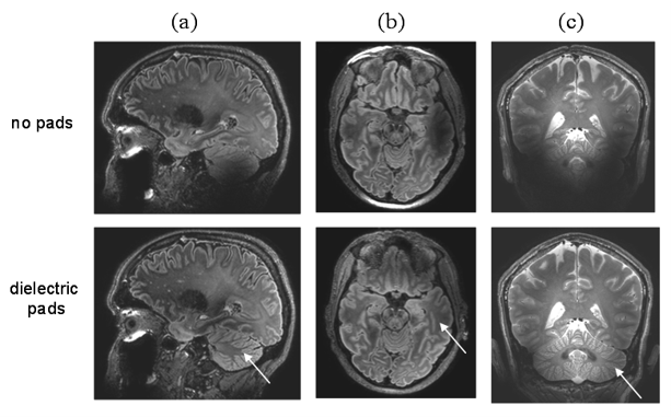
High field MRI holds great promise for many clinical and clinical research applications. However, there are also a large number of technical challenges which need to be overcome to take full advantage of the high magnetic field strength. Our group develops novel solutions to these challenges, including new high permittivity materials and artificial dielectrics to improve image uniformity, as well as RF transmitters which reduce the amount of power deposited in the patient. We have also translated these approaches to clinical field strengths of 1.5 and 3 Tesla.
…High field MRI holds great promise for many clinical and clinical research applications. However, there are also a large number of technical challenges which need to be overcome to take full advantage of the high magnetic field strength. Our group develops novel solutions to these challenges, including new high permittivity materials and artificial dielectrics to improve image uniformity, as well as RF transmitters which reduce the amount of power deposited in the patient. We have also translated these approaches to clinical field strengths of 1.5 and 3 Tesla.
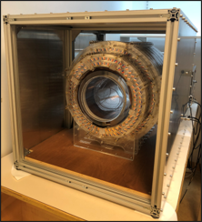
Globally over 70% of the world’s population has absolutely no access to MRI, and clinical conditions which could benefit from even very simple scans cannot be treated. Rather than designing a highly sophisticated and expensive piece of equipment that can be used for all types of scanning, we use the philosophy of tailored design, such that we can design much more inexpensive systems for specific medical applications. we design systems that use thousands of very small low-cost permanent magnets, arranged in designs which have no fringe field and therefore very easy siting requirements. The low magnetic fields allow scanning of patients with implants, and the scanner could potentially be transported on an ambulance for differentiation of hemorrhagic or ischemic stroke.
…Globally over 70% of the world’s population has absolutely no access to MRI, and clinical conditions which could benefit from even very simple scans cannot be treated. Rather than designing a highly sophisticated and expensive piece of equipment that can be used for all types of scanning, we use the philosophy of tailored design, such that we can design much more inexpensive systems for specific medical applications. we design systems that use thousands of very small low-cost permanent magnets, arranged in designs which have no fringe field and therefore very easy siting requirements. The low magnetic fields allow scanning of patients with implants, and the scanner could potentially be transported on an ambulance for differentiation of hemorrhagic or ischemic stroke.
Projects
Themes for Innovation
Key publications
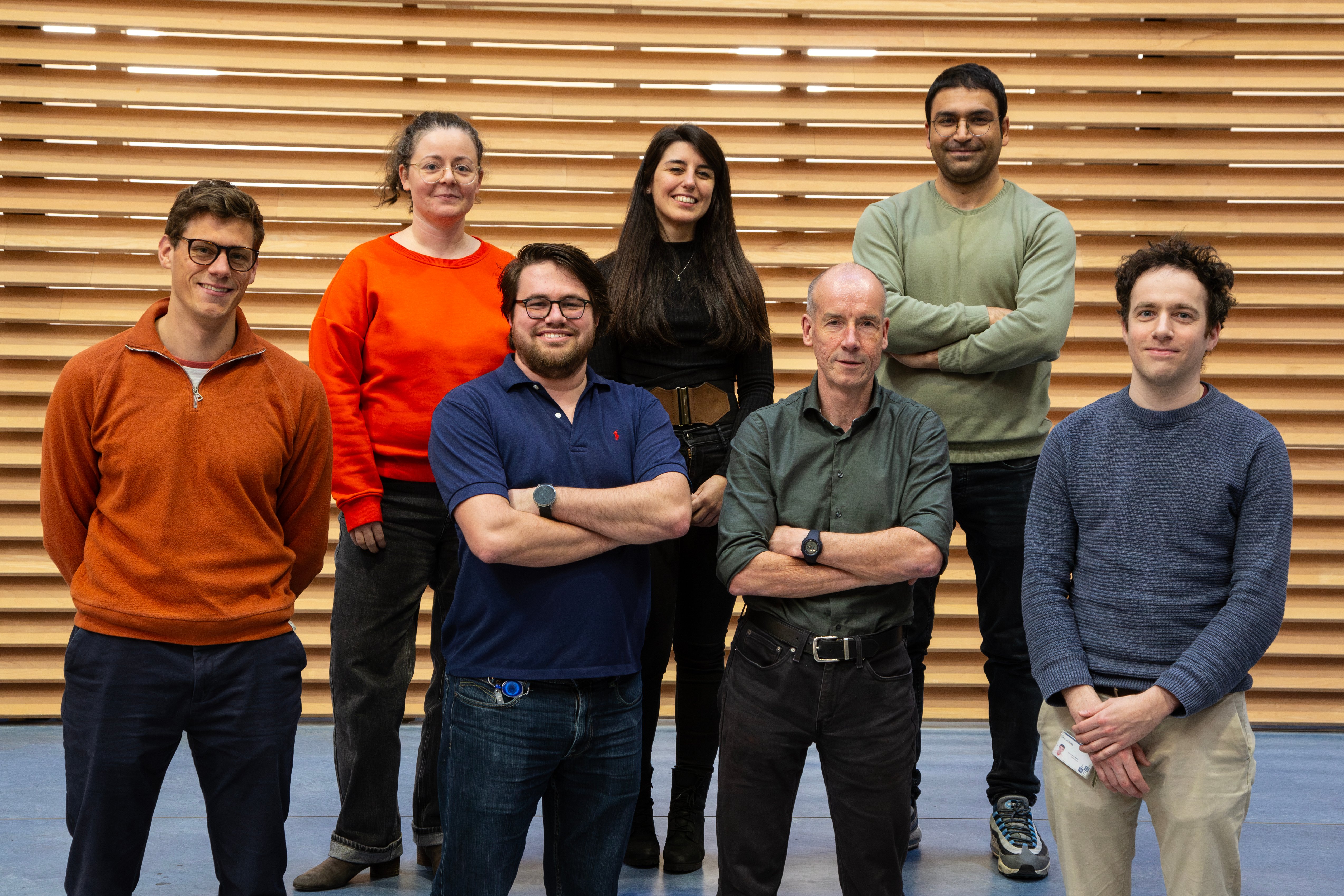
Team members
Current members:
- Andrew Webb, PI
- Beatrice Lena, postdoctoral researcher
- Chloe Najac, postdoctoral researcher
- Tom O'Reilly, postdoctoral researcher
- Aram Salehi, PhD candidate
- Bart de Vos, PhD candidate
- Javad Parsa, PhD candidate
- Ruben van der Broek, clinical technologist
- Jan Groen, Philips Associate
Former members:
- Wouter Teeuwisse, senior scientist
- Mathieu Mach, Research assistent
- Wyger Brink, postdoctoral researcher
- Reijer Leijsen, PhD candidate
- Wico Breimer, PhD candidate
- Jelle Hockx, PhD candidate